Petri Dish under the Microscope
Channel: Sci- Inspi
Category: Science & Technology
Tags: myceliumbiologybacteriamicrobiologymoldpetri dishmicroscopeguitaramazingfungussciencehyphaepetri plate
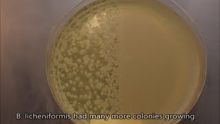
Description: Microscopic footage of colonies from a petri plate that a student inoculated. The student touched this petri plate against a bench in one of our chemistry labs. He left it to incubate at room temperature for about a week. The medium used is D/E neutralizing agar. The smooth colonies are most likely bacterial while the hairy colonies are fungal molds. Mold gets its furry look from hyphae, which are long thread like filaments which release enzymes and absorb nutrients. Colonies change over time. This video shows what they look like after 4 days. Camera: Nikon D3300 Microscope: Wolfe Steromicroscope - 10X, 30X Magnification of each shot is shown at the bottom right hand corner.