Sleep Physiology, Animation
Channel: Alila Medical Media
Category: Education
Tags: yt:quality=high
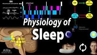
Description: Stages of sleep, REM and NREM sleep, mechanism of regulation, sleep- and wake-promoting regions (VLPO, TMN and hypocretin neurons) of the brain, homeostatic drive and circadian rhythm. This video is available for instant download licensing here: alilamedicalmedia.com/-/galleries/all-animations/brain-and-nervous-system-videos/-/medias/c13647eb-b374-416f-a48d-a8f51aef5dd3-sleep-physiology-narrated-animation ©Alila Medical Media. All rights reserved. Voice by : Marty Henne Support us on Patreon and get early access to videos and free image downloads: patreon.com/AlilaMedicalMedia All images/videos by Alila Medical Media are for information purposes ONLY and are NOT intended to replace professional medical advice, diagnosis or treatment. Always seek the advice of a qualified healthcare provider with any questions you may have regarding a medical condition. Sleep is a temporary state of unconsciousness in which the brain is primarily responsive to internal, rather than external stimuli. Unlike other states of unconsciousness such as coma or general anesthesia, sleep is a natural, cyclic process that is self-regulated and easily reversible to wakefulness. Brain activity can be recorded in the form of electroencephalogram, EEG, which measures electrical activities in the superficial layers of the cerebral cortex. Different stages of consciousness correspond to different types of brain waves. A fully awake and alert brain produces high-frequency low-voltage beta-waves. As consciousness decreases, brain waves become progressively slower in frequency and higher in voltage. There are 2 major phases of sleep: rapid eye movement, REM, sleep, and non-rapid eye movement, non-REM, sleep. Non-REM sleep progresses in 3 stages: N1, N2 and N3 N1 is the transitional state between wakefulness and sleep. The EEG is dominated by alpha-waves. The sleeper is easily awoken with light stimulation. N1 typically lasts a few minutes. The next stage is N2, a deeper sleep state, where stronger stimuli are required to produce awakening. Brain activity is slower and more irregular, with short bursts of “sleep spindles” and “K-complexes.” It is believed that memory consolidation occurs during this stage. N3 is deeper than N2. Slow delta-waves dominate. Muscles relax, vital signs are at their lowest; and it is difficult to wake the sleeper. N3 is typically followed by a transition to N2 before REM sleep occurs. As its name suggests, REM sleep is characterized by rapid eye movements under the eyelids. It’s also known as “paradoxical” sleep because the brain’s EEG is very much similar to that of the waking state. REM sleep is when most dreams occur, as well as some autonomic reflexes. Vital signs are up, but there is a total inhibition of skeletal muscles, which prevents sleepers from acting out their dreams. This sequence of stages repeats itself 4 to 5 times in a typical night. As the night progresses, the duration of N2 and REM sleep increases, while N3 decreases. The amount and timing of sleep is regulated by 2 major factors: homeostatic drive and circadian rhythm. Homeostatic drive is basically the body’s need for sleep, or pressure to sleep. Adenosine is thought to be a substance that accumulates with waking hours and drives the pressure to sleep. Interestingly, caffeine appears to promote wakefulness by acting as an antagonist of adenosine. The need to sleep increases with illness, as well as cognitively stimulating or physically demanding activities. Circadian rhythm is the body’s biological clock for the sleep-wake cycle. It determines the timing of sleep. The master clock is located in the suprachiasmatic nucleus, the SCN, of the hypothalamus. It receives light inputs from the retina and resets the clock everyday accordingly to the day-night cycle. The SCN is most active during the day, and least active at night. The sleep-promoting region is located in the ventrolateral preoptic nucleus, VLPO, of the hypothalamus. The VLPO is inhibited by the SCN and activated by adenosine. The VLPO uses GABA to inhibit wake-promoting regions of the brain, which include multiple nuclei in the reticular formation and posterior hypothalamus. Of these regions, it’s important to note the tuberomammillary nucleus, TMN, and the hypocretin neurons. The TMN consists mainly of histaminergic neurons, but it also produces GABA that inhibits VLPO in return. This mutual inhibition is the basis of the “switch” between sleep and wake. The hypocretin neurons stimulate the TMN, and are crucial for maintaining wakefulness. The loss of these neurons results in narcolepsy. There is a similar switch between REM and non-REM sleep, mediated by mutually inhibiting REM-on and REM-off neurons in the pons.